

Intraoral scanners (IOS) have become an important part of digital dentistry, providing digital impressions for restorative, orthodontic, and implant workflows. However, the accuracy of an intraoral scan is not determined by the scanner alone.
The final digital model is the result of a complete process involving optical acquisition, image processing, data registration, 3D reconstruction, scanning technique, and the condition of the oral environment.
Understanding these factors is important when evaluating why an intraoral scan may be accurate in one situation but less consistent in another.
What Does Intraoral Scanner Accuracy Mean?
Before discussing the influencing factors, it is useful to distinguish between two concepts: trueness and precision.
Trueness
Trueness describes how closely the digital scan matches the actual geometry of the object being scanned.
In simple terms:
Actual geometry → Digital model → Geometric deviation
A smaller deviation generally indicates higher trueness.
Precision
Precision describes how consistently repeated scans produce similar results.
For example, if the same dental arch is scanned several times and the resulting digital models are highly similar, the scanning process demonstrates good precision.
These two concepts are related but not identical.
A scanner may repeatedly produce very similar results while still having a systematic deviation from the actual geometry.
Therefore:
High precision does not automatically mean high trueness.
Both characteristics are important when evaluating intraoral scanning accuracy.
1. Optical System Design
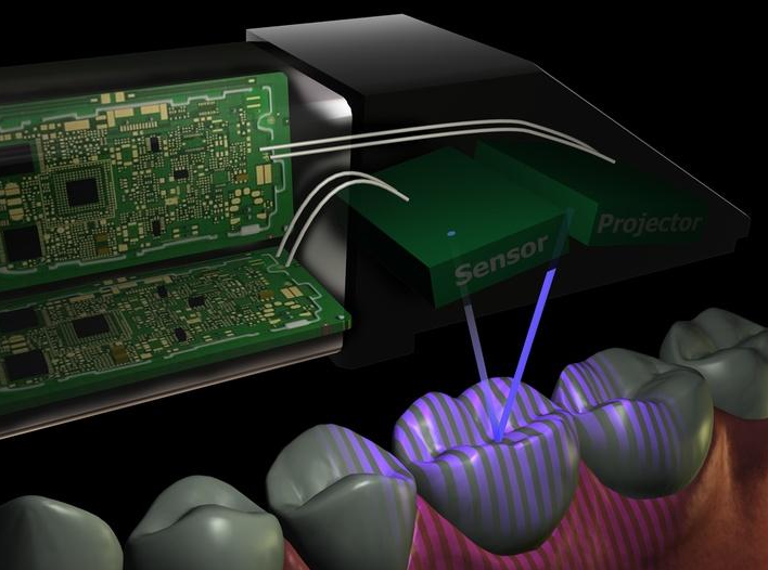
The optical system is the starting point of the scanning process.
Inside the scan head, components such as:
- Light sources
- Lenses
- Image sensors
- Mirrors or prisms
- Projection optics
- Protective windows
work together to capture information from the oral surface.
Different optical architectures can use different approaches, including direct optical paths, folded optical paths, multi-view acquisition, projection-based imaging, or confocal principles.
The optical design affects the quality of the raw information available for 3D reconstruction.
A simplified process is:
Optical system → Image acquisition → Data processing → 3D reconstruction
If the original optical information is incomplete or unstable, software processing cannot necessarily compensate for every limitation.
Field of View and Depth of Field
Two important optical characteristics are the field of view and depth of field.
Field of view determines how much of the oral surface can be captured from a particular position.
Depth of field describes the range of distances over which useful image information can be maintained.
These characteristics influence how easily the scanner can capture curved, deep, or narrow anatomical structures.
2. Scan Head Condition and Optical Window
The protective window at the front of the scan head separates the internal optical system from the oral environment.
Because light must pass through this window, its condition can directly affect image acquisition.
Potential problems include:
- Fogging
- Saliva contamination
- Water droplets
- Residue
- Scratches
- Surface contamination
Why Does Fogging Matter?
Fogging changes the optical conditions between the scanner and the oral surface.
It can introduce scattering or reduce the clarity of the captured information, making it more difficult for the imaging system to obtain stable data.
Therefore, anti-fogging is not simply a matter of operator comfort. Maintaining a clear optical window can help preserve consistent image acquisition.
3. Optical Properties of the Scanning Surface
Teeth and dental materials are not ideal optical surfaces.
Their optical behavior can vary according to:
- Reflectivity
- Translucency
- Surface texture
- Gloss
- Color
- Moisture
Highly Reflective Surfaces
Strong reflections can create areas where the captured optical information is less stable.
Translucent Surfaces
Light may interact with translucent materials differently from opaque surfaces, potentially creating more complex optical signals.
Smooth Surfaces
Very smooth surfaces may contain fewer distinctive geometric or textural features for image registration.
This is why scanner performance cannot be evaluated only by asking how high the camera resolution is.
The interaction between the optical system and the scanned surface also matters.
4. Saliva, Blood, and Moisture
The oral cavity is a challenging scanning environment.
Saliva and other fluids can change the optical properties of the surface being scanned.
They may cause:
- Specular reflections
- Local obstruction
- Reduced image contrast
- Changes in surface appearance
- Temporary loss of visible features
Blood or other contamination can create additional visual interference.
For this reason, controlling the scanning field is an important part of obtaining consistent digital impressions.
Keeping the target area as clean and appropriately dry as the clinical situation allows can make the optical information more stable.
5. Scanning Distance and Angle
The distance and angle between the scan head and the tooth surface also affect image acquisition.
Scanning Distance
Every optical system operates within a particular working range.
If the scan head moves too far away or too close to the target surface, image quality may decrease.
Possible effects include:
- Reduced focus
- Changes in effective field of view
- Loss of useful surface information
Scanning Angle
The scanner also needs an appropriate viewing angle to capture different surfaces.
An extreme angle can make certain areas difficult to observe, especially around:
- Interproximal surfaces
- Lingual surfaces
- Deep anatomical structures
- Posterior regions
This is one reason why the operator’s scanning technique remains important even when the scanner itself has advanced optical hardware.
6. Scanning Path and Scanning Strategy
An intraoral scanner does not simply take one photograph of the entire dental arch.
Instead, it continuously captures image data and combines those datasets into a larger 3D model.
A simplified process looks like:
Frame 1 → Frame 2 → Frame 3 → Frame 4 → … → 3D model
The software must determine how each newly captured frame relates to the previously acquired data.
Therefore, scanning strategy can influence the stability of data registration.
Potential challenges include:
- Abrupt changes in scanning direction
- Excessively long continuous scanning paths
- Repeated scanning of similar surfaces
- Spending too much time in areas with limited geometric features
- Moving away from previously captured reference information
A well-planned scanning path can provide the software with more reliable information for continuous registration.
7. Surface Features and Data Registration
Data registration is one of the key steps in converting a sequence of images into a coherent 3D model.
The software needs to identify corresponding information between consecutive datasets.
For example:
New frame → Identify common features → Estimate spatial relationship → Align with existing data
If the scanned surface contains sufficient distinctive features, registration can generally be more reliable.
However, large areas with repetitive or relatively featureless geometry can make matching more challenging.
This becomes particularly important during full-arch scanning, where a small registration error may potentially propagate as the scanning sequence becomes longer.
Does a Longer Scan Always Mean More Error?
Not necessarily.
A larger scanning range creates more opportunities for registration challenges, but the final accuracy depends on many factors, including:
- Optical quality
- Surface characteristics
- Scanning strategy
- Registration algorithms
- Calibration
- Operator technique
Therefore, it would be inaccurate to assume that every longer scan automatically produces proportionally larger errors.
8. Geometric Complexity of the Scanning Area
Different anatomical regions present different scanning challenges.
For example:
- Deep grooves
- Interproximal areas
- Gingival margins
- Distal surfaces of posterior teeth
- Narrow spaces
- Complex restoration margins
may require different viewing angles and scanning movements.
The difficulty of scanning therefore depends partly on the geometry of the target itself.
A useful way to understand this relationship is:
Scanner capability + target geometry + scanning technique
All three interact to determine the quality of the final digital model.
9. Scanner Calibration
Calibration establishes the geometric relationship between the optical components and the measurements generated by the system.
If the relative position of optical components changes, or if calibration parameters no longer accurately represent the system, measurement accuracy can be affected.
Calibration may account for factors related to:
- Optical geometry
- Sensor position
- Camera parameters
- System geometry
- Manufacturing tolerances
The purpose is not simply to “make the scanner more accurate.”
Rather, calibration creates a reliable relationship between the captured optical information and the corresponding 3D geometry.
10. Temperature and System Stability
An intraoral scanner is a precision system containing optical, electronic, and mechanical components.
Temperature changes can potentially affect:
- Optical components
- Image sensors
- Electronic components
- Mechanical structures
Even small physical changes can influence the relative geometry of a precision optical system.
Depending on the system design, temperature-related effects may be addressed through measures such as:
- Thermal stabilization
- Warm-up procedures
- Calibration
- Software compensation
However, software compensation should not be viewed as a universal solution.
It needs to be based on a valid measurement model and appropriate calibration data.
11. Operator Technique
Even with sophisticated optical hardware and software, the operator remains an important part of the scanning process.
Operator-related factors include:
- Scanning speed
- Scanning distance
- Scanning angle
- Movement stability
- Scanning path
- Management of saliva and contamination
- Re-scanning of missing areas
Two operators using the same scanner may therefore obtain different scanning results if their scanning techniques differ.
This does not necessarily mean that the scanner itself is inaccurate.
Instead, it demonstrates that intraoral scanning is a system-level process involving both technology and operation.
12. Data Processing and 3D Reconstruction
After optical information is captured, it still needs to be converted into a usable digital model.
A simplified workflow is:
Image acquisition → Feature extraction → Registration → Alignment → Data processing → 3D reconstruction
Each step can influence the final result.
For example, registration determines how different datasets are spatially connected, while reconstruction converts the processed information into a continuous digital surface.
This leads to an important distinction:
Optical structure and scanning algorithm are not the same thing.
The optical system determines how physical information is captured.
The software determines how that information is interpreted, aligned, processed, and reconstructed.
Both are necessary for accurate digital scanning.
13. Why Full-Arch Scanning Can Be More Challenging
Scanning a single tooth and scanning an entire dental arch are not equivalent tasks.
A single-tooth scan covers a relatively small area.
A full-arch scan requires the system to maintain reliable spatial relationships across a much larger dataset.
The workflow may involve:
Local acquisition → Continuous registration → Error control → Global alignment → Full-arch reconstruction
This is why full-arch scanning can place greater demands on:
- Optical stability
- Surface feature recognition
- Registration algorithms
- Calibration
- Scanning strategy
- Operator technique
However, full-arch accuracy should not be reduced to a single factor. It is the combined result of the entire scanning system.
How the Factors Work Together
The accuracy of an intraoral scan is best understood as a chain rather than a single specification.
Optical Design
↓
Raw Image Quality
↓
Surface Information
↓
Feature Extraction
↓
Data Registration
↓
Error Control
↓
3D Reconstruction
↓
Final Digital Model
A limitation at one stage may affect the stages that follow.
At the same time, improvements in one component do not necessarily eliminate limitations elsewhere.
For example, a high-quality image sensor cannot completely solve poor scan-head optics, while sophisticated software cannot always recover information that was never captured.
Conclusion
The accuracy of an intraoral scanner is influenced by much more than the scanner’s advertised specifications.
The main factors can be grouped into five areas:
- Scanner hardware — optical design, sensors, scan-head structure, and calibration.
- Optical environment — reflections, moisture, fogging, and surface optical properties.
- Scanning target — surface texture, geometry, visibility, and material characteristics.
- Scanning technique — distance, angle, speed, and scanning path.
- Data processing — registration, alignment, error control, and 3D reconstruction.
Ultimately, intraoral scanning accuracy is the result of the interaction between optics, mechanics, sensors, calibration, software, the scanning environment, and operator technique.
Post time: Sep-30-2026